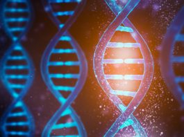
Researchers at universities in Yorkshire have developed videos based on high-resolution images of a DNA helix, allowing us to see ‘dancing DNA’ for the first time.
Researchers from the Universities of Sheffield, York and Leeds have captured the highest resolution images of a single DNA helix.
Studies revealed that the stresses and strains placed on DNA when it is packed inside cells, can lead to it changing shape; the DNA helix adopts dance like movements, making twisting and turning movements inside a cell.
The high-resolution images, combined with supercomputer simulations, have allowed scientists to develop video footage of the position of every single atom in the DNA and how it twists, allowing us to see the ‘dancing DNA’ for the first time.
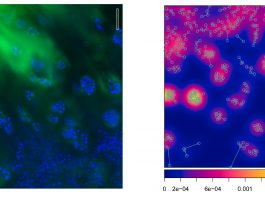
The images are so detailed that it is possible to see the double helical structure of the DNA, but when combined with the computer simulations, the researchers were able to see the position of every single atom in the DNA and how it moves.
Every cell in the human body contains two metres of DNA, so for it to fit inside the cells, it has evolved to twist and turn. Loopy DNA is therefore everywhere in the genome, forming twisted structures.
The team looked at DNA minicircles, where the molecule is joined at both ends to form a loop. This loop allowed the researchers to give the DNA minicircles an added twist, causing it to ‘dance’ more vigorously.
When they imaged relaxed DNA, without twists, it did not do much. However, when they gave the DNA an added twist, it became far more dynamic and adopted dance like shapes. These dance-moves were found to be the key to finding binding partners for the DNA, as when they adopt a wider range of shapes, then a greater variety of other molecules find it attractive.
DNA minicircles in cells can twist and bend and become very compact. Being able to study DNA at such detail could help lead to advancement of new gene therapies by utilising the way compacted DNA circles can squeeze their way into cells.
Dr Alice Pyne, lecturer in Polymers & Soft Matter at the University of Sheffield, who captured the footage, said: “Seeing is believing, but with something as small as DNA, seeing the helical structure of the entire DNA molecule was extremely challenging.
“The videos we have developed enable us to observe DNA twisting in a level of detail that has never been seen before.”
Dr. Agnes Noy, EPSRC Early Career Fellow and Lecturer at the University of York, who did the theoretical modelling in the study, said: “The computer simulations and microscopy images agree so well that they boost the resolution of experiments and enable us to track how each atom of the double helix of DNA dances.”