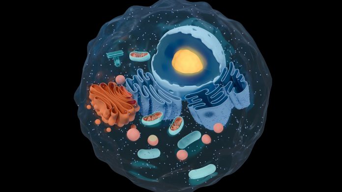
Researchers at the University of Warwick have discovered a protein that bends the cytoskeleton of cells, twisting them into novel shapes.
This unique cell cytoskeleton twisting protein, known as the ‘curly’ protein, may play a fundamental role in cell division, a process vital to all living matter. As well as this, it offers biologists an exciting new engineering tool that can be utilised to modify cell shapes.
The ‘curly’ protein
According to the Warwick researchers, a protein that causes a cell’s cytoskeleton to bend, thus enabling the cell to twist into various shapes, may be key to the process of cell division.
By causing the cytoskeleton to bend inwardly to a tight point at its core, one cell divides into two – much like how you would make a balloon animal by twisting. This ‘curly’ protein has been detected twisting the material that comprises the cytoskeleton for the first time.
The team’s results, which have been obtained from research supported by the European Research Council and Wellcome Trust, have been published in the journal eLife, and indicate novel and exciting ways that cells can be modified and engineered. On top of this, the results could lead to a greater understanding of how they replicate – a process vital to all living organisms.
Understanding cell division
All living matter is made up of cells, which are encompassed by a delicate, yet malleable membrane similar to a balloon. This is reinforced by a cytoskeleton, made up of a material called actin, that provides shape and stability to cells. Filaments of actin are semi-flexible and generally straight, and it has long been believed that they establish structures by stacking together like matchsticks.
Throughout the process of cell division, a cell will develop a cytokinetic ring, which is a type of machinery comprised of actin and motor proteins called myosin, along the centre of the surrounding membrane. The myosins then constrict the actin filaments and squeeze the cell down its middle until it separates.
Until now, scientists believed that the actin filaments that form the cytokinetic ring split up as the cell is squeezed tighter. An interdisciplinary partnership between Professor Mohan Balasubramanian, Dr Saravanan Palani (now at the Indian Institute of Science) and Dr Darius Köster at the University of Warwick was, therefore, amazed to discover that a segment of the protein Rng2, nicknamed ‘curly’, can naturally bend actin, with 10 micrometres of actin creating a ring less than a micrometre in diameter.
This indicates that ‘curly’ could play a significant role in enabling the cytokinetic ring to shrink tightly enough to divide a cell.
Balasubramanian, one of the leading researchers, discovered Rng2 – an important protein in this process – over two decades ago, but only now have the researchers been able to make the fundamental observation that the protein can bend actin filaments into rings.
Rng2 is related to the highly conserved IQGAP family of proteins that are present in an array of things, from yeast to humans, so the results of this research could be applicable to other cells as well, including human cells.
Köster, of Warwick Medical School, said: “What we show is if we have this protein on the membrane and we polymerise fresh actin filaments at the membrane, representing what is happening in the cytokinetic ring, the actin starts to bend. If you have it in a cell, it’s a curved surface, so the actin would curve.
“As far as we know, there are no other proteins doing this kind of job with individual filaments. If you look inside cells, there are definitely curved actin structures, but it was thought that they were stacks of very short straight filaments packed together. This opens the perspective that it could be geometries and certain organelles that the actin is actually wound around.”
The reconstitution approach
The team employed something called a reconstitution approach, whereby they investigate an intricate system by dividing it into its constituent parts, then reconstruct it to decipher which role each individual element plays.
For the research, the group produced a lipid bilayer to characterise the cell membrane and experimented with adding various proteins to it that are typically located at the interface with the cytokinetic ring during cell division.
They then added actin filaments and myosin to represent the ring itself. Through this, they discovered that when ‘curly’ was present, the actin formed highly curved structures, which is behaviour not normally observed in these kinds of experiments.
This opens the prospect of utilising ‘curly’ as a tool to engineer various shapes of cells. Scientists could potentially use it to alter the cytoskeleton and twist the cell into new shapes.
Dr Köster added: “We have very few tools we can use to modify the cell cortex, and for the first time, we have a way to engineer bent actin structures. Since we can express it in cells and the cells react to it, we can use it to modify what the cell cortex looks like.
“This can help us to really understand how curved actin can deform membranes, or whether the cell uses this as a mechanism to control certain processes. There seem to be some proteins that like to twist and change the curvature of actin, and if that’s happening, then it’s very likely that there will be other proteins that take that as an information cue.”


