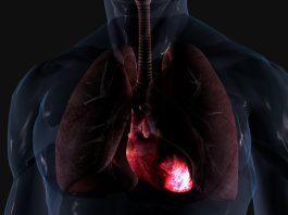
The development of a novel robot has potentially changed the landscape of future cardiac surgery, mitigating fatigue and radiation exposure for human operators.
Invented by a team of international researchers, the innovative technology provides efficient robot-assisted cardiac surgery that enables cardiac surgeons to perform in-depth planning of operations and enhances the surgical field of view. The robot is operated by a virtual reality parallel system as a digital twin, proficient in accurately conducting ultrasound imaging of a patient whilst mitigating detrimental effects caused by the procedure to the medical professional operating it.
Their research is published in IEEE/CAA Journal of Automatica Sinica.
Fei-Yue Wang, the author of the study and Director of the State Key Laboratory of Management and Control of Complex Systems, Institute of Automation, Chinese Academy of Sciences, said: “Intra-operative ultrasound is especially useful, as it can guide the surgery by providing real-time images of otherwise hidden devices and anatomy. However, the need for highly specialised skills is always a barrier for reliable and repeatable acquisition.”
The presence of onsite sonographers can be quite low in medical facilities, with various procedures requiring intra-operative ultrasound that potentially utilises X-ray imaging, potentially putting operators at risk through harmful radiation. To overcome this hazardous obstacle, the team fabricated a platform for robotic intra-operative trans-oesophagal echocardiography (TEE), a technique employed to diagnose heart disease and assist cardiac surgery.
Wang said: “Our result has indicated that the use of the robot with a simulation platform could potentially improve the general usability of intra-operative ultrasound and assist operators with less experience.”
The new technology consists of parallel control and intelligence that pairs the operator and robot in a virtual environment which accurately mirrors the real environment; it is equipped with an extensive database of ultrasound images in addition to a digital platform proficient in reconstructing anatomy. The robot is able to navigate specific target areas for the operator, allowing them to visualise and plan potential surgical corrections in computational experiments accurately.
“Such a system can be used for view definition and optimisation to assist pre-planning, as well as algorithm evaluations to facilitate control and navigation in real-time,” Wang added.
The researchers are now aiming to integrate the parallel real/virtual system with the specialised clinical need to aid in the translational research of imaging robots.
“The ultimate goal is to integrate the virtual system and the physical robot for in-vivo clinical tests, so as to propose a new diagnosis and treatment protocol using parallel intelligence in medical operations,” Wang commented.