From wearables to implants, graphene and other layered materials have great potential for numerous next-generation biomedical technologies. Here, Letizia Diamante, Science Communicator at the Graphene Flagship, discusses more.
The rapidly evolving field of graphene-based technologies for biomedical applications holds tremendous promise to offer improved treatments, diagnostics, and monitoring methods for a variety of diseases.
Graphene exhibits extraordinary characteristics that render it a highly desirable material for diverse applications in the field of medicine and healthcare. Its exceptional strength, flexibility, electrical conductivity, biocompatibility, transparency, and large surface area have opened up avenues for advancements in various domains, including biosensors and diagnostic tools. One notable example is graphene’s ability to interact with electro-active cells and tissues, which holds great promise for medical monitoring.
In this article, we will explore some of the latest developments in graphene-based biomedical technologies and their potential impact on the future of healthcare.
The European spin-off pioneering implantable neural platform made of graphene
Existing neural interfaces for brain implants are based on metals, such as platinum and iridium, which impose significant restrictions in terms of miniaturisation, signal resolution, side effects and high rejection rate in patients. The spin-off company and Partner of the Graphene Flagship, INBRAIN Neuroelectronics, which originated from Catalan Institute of Nanoscience and Nanotechnology (ICN2) and ICREA in Spain, is developing implants based on graphene for decoding and modulating neural activity. The spin-off already attracted more than €25m in capital and non-dilutive investments, and secured a collaboration with the pharma company Merck KGaA. Its highly promising graphene interfaces, combined with advanced electronics and data analysis provided by Machine Learning and Deep Learning, promise to decode neural signals at high resolution from both the central and peripheral nervous systems.
Graphene-based biomedical technologies offer numerous benefits in comparison to existing platinum-based solutions. Firstly, graphene is remarkably soft and highly flexible, akin to an ‘electronic skin’, allowing for optimal contact with the brain surface. Additionally, graphene enables the development of miniaturised brain sensors, up to 10,000 times smaller than platinum-based sensors. This reduces invasiveness and could help prevent unwanted side effects.
The real-time, high resolution of INBRAIN’s systems is another major advantage. Graphene devices offer up to 1,024 sensing dots, allowing for the decoding of neural signals in both high density and ultra-high resolution. This capability extends to frequency ranges that metals cannot reach, thereby uncovering new neural biomarkers.
The biocompatibility and toxicity of INBRAIN’s technology has been tested in vitro and in vivo. It has been used successfully in studies on in-silico and animal models, and clinical trials are expected to start soon. Ultimately, a longer-term goal for INBRAIN engineers is to explore how graphene-based biomedical technologies can decode neuronal circuits to rehabilitate patients suffering from central nervous system conditions, such as Parkinson’s, epilepsy, and speech impairments, but also peripheral nerve conditions, such as rheumatoid arthritis. The vision is to fully decode and modulate the entire nervous system, illuminating a path towards a better neural future for the one third of the population suffering from neural-related disorders.
Monitoring slow brain waves, known as brain tsunamis
Just as tsunamis are colossal waves that surge across the ocean, spreading depolarisation (or brain tsunamis) can sweep through the brain, causing disruption to its normal functioning. These abnormal brain activities can be triggered by traumatic brain injuries, strokes, and other neurological diseases. During spreading depolarisation, the delicate balance of signals that allow neuron-to-neuron communication is disrupted, impairing the neurons’ ability to function properly.
Monitoring spreading depolarisations is complicated, and standard electroencephalography (EEG) may not effectively capture or differentiate them. These slow waves have frequencies below 0.1 Hz and ongoing research is still unravelling their precise functions and clinical implications. Their frequency is significantly lower compared to other brain waves, such as theta waves occurring during light sleep and delta waves that are typical of deep sleep.
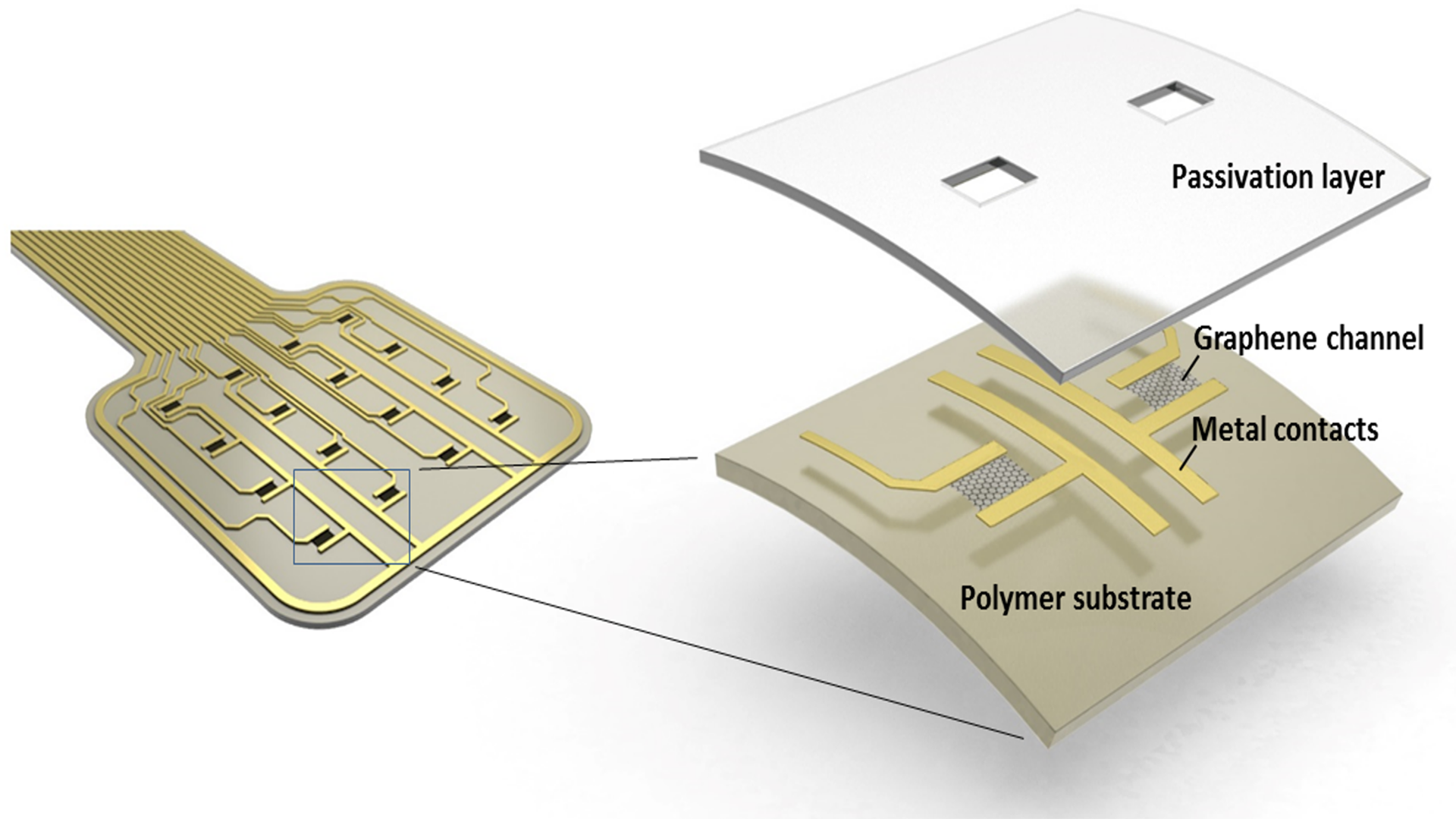
Glass microelectrodes, commonly referred to as pipettes, have traditionally served as a fundamental tool in the field of electrophysiology. However, their utility is limited to measuring brain electrical signals at specific points. In an effort to expand the coverage of brain activity, a team of researchers associated with the Graphene Flagship project has developed flexible graphene field-effect transistors (FETs) and depth neural probes. These innovative graphene depth neural probes have been utilised to investigate the brain activity of awake rodents affected by epilepsy. Implanting these graphene-based biomedical technologies in rats for a period of ten weeks has facilitated the recording of brain activity and the associated electrophysiological markers of epilepsy, including spreading depolarisations. This breakthrough offers promising insights into understanding and monitoring brain disorders, showcasing the potential of graphene-based technologies in advancing the field of neuroscience.1,2,3
This breakthrough paves the way for future clinical applications in pre-surgical monitoring and neurological care to enhance the identification of seizure onset zones and improved surgical outcomes. Looking beyond epilepsy research, the researchers believe this technology holds promise for advancing our understanding of other neurological disorders associated with spreading depolarisation, including traumatic brain injury, stroke, and migraine.
Diagnosing eye conditions with graphene electrodes
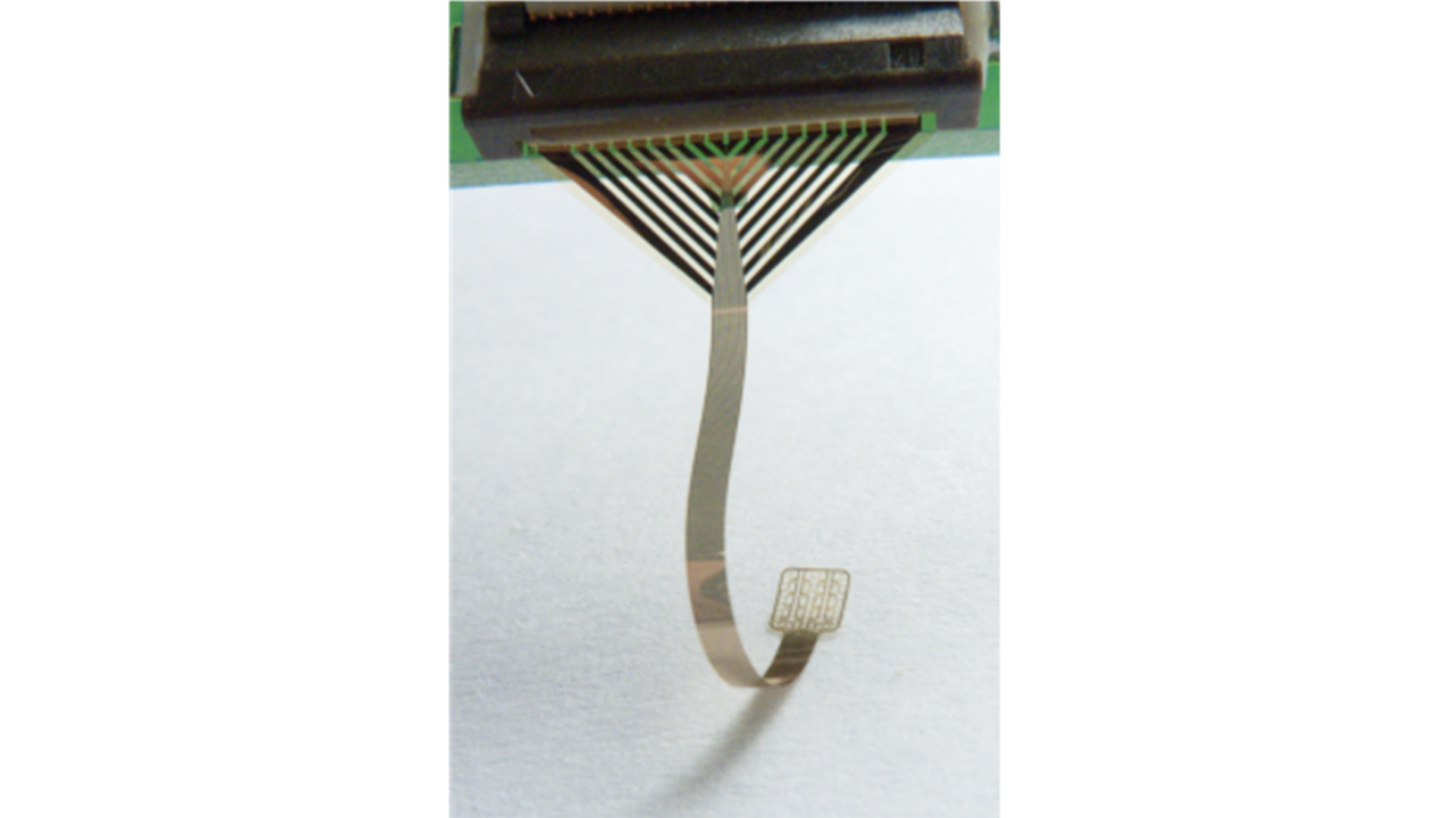
Research teams led by Serge Picaud of Sorbonne University (France) and Jose A Garrido of ICN2 carried out electroretinography – a diagnostic test that measures the electrical activity of the retina to diagnose various eye conditions – with graphene electrodes.
Typically, electroretinography involves the placement of a gold wire electrode on the cornea, followed by the stimulation of the eye with a flashing light to measure changes in the transretinal potential. However, these electrodes lack a flexible interface to effectively contact the delicate corneal tissue, resulting in discomfort for the patient. Furthermore, their opacity restricts the recording of signals coming from the corneal periphery.
To overcome these challenges, the researchers have developed flexible electrodes using graphene-based biomedical technologies. Leveraging the transparency of graphene, the team fabricated microelectrode arrays and tested them on rats. The results demonstrated the superior performance of graphene-based electrodes compared to the traditional gold electrodes.
This innovative application of graphene in electrodes to record electrical signals directly on the cornea could provide enhanced comfort for patients, improve recording capabilities, and capture signals coming from the centre of the cornea.4
Using graphene-based biomedical technologies for biosensors in smart patches and quick screenings
In the ever-expanding realm of wearables, researchers of Graphene Flagship Associate Member Grapheal have developed WoundLAB – a patch to monitor the pH and other parameters of wounds. The flexible and transparent graphene-based biosensor continuously records and stores data about the wound, which is then communicated to the cloud thanks to a smartphone app. Using this technology, doctors and nurses can remotely monitor wound healing and check if any infections or medical complications arise.
Grapheal also won the prestigious CES 2022 Innovation Award in Las Vegas for TestNpass – a battery-free device that uses graphene-based biomedical technologies to rapidly screen for infection from liquids (e.g. saliva samples). While lateral flow tests to screen for COVID-19 take about 20 minutes, TestNpass shows the result in fewer than three minutes. Furthermore, it takes advantage of smartphones’ contactless communication technology, called NFC, to show the results directly in an app.
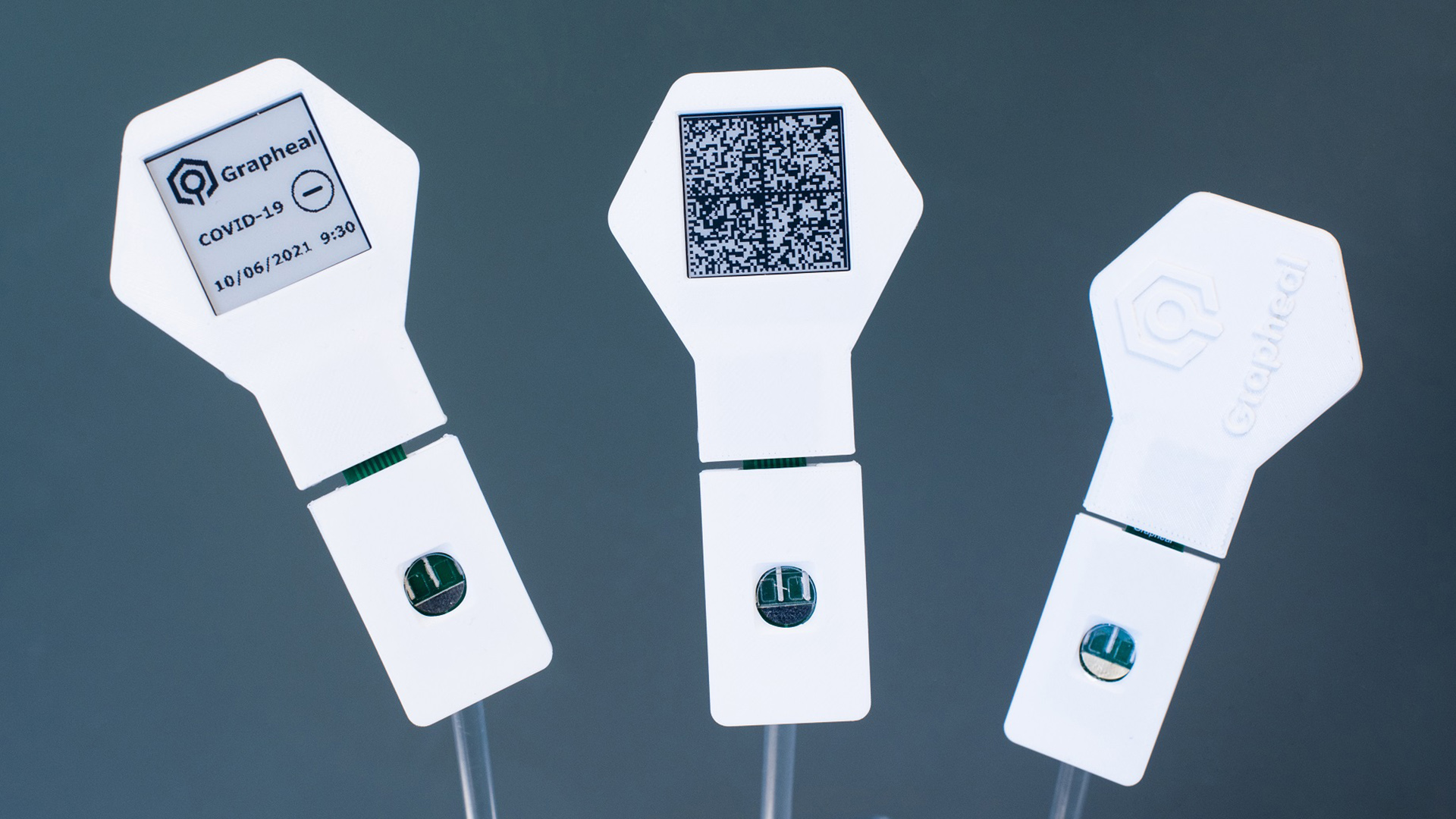
The company envisions functionalisation methods to anchor graphene to other proteins and antibodies, specific to different pathogens and diseases.
Developing an automated cancer detection system with graphene-based technologies
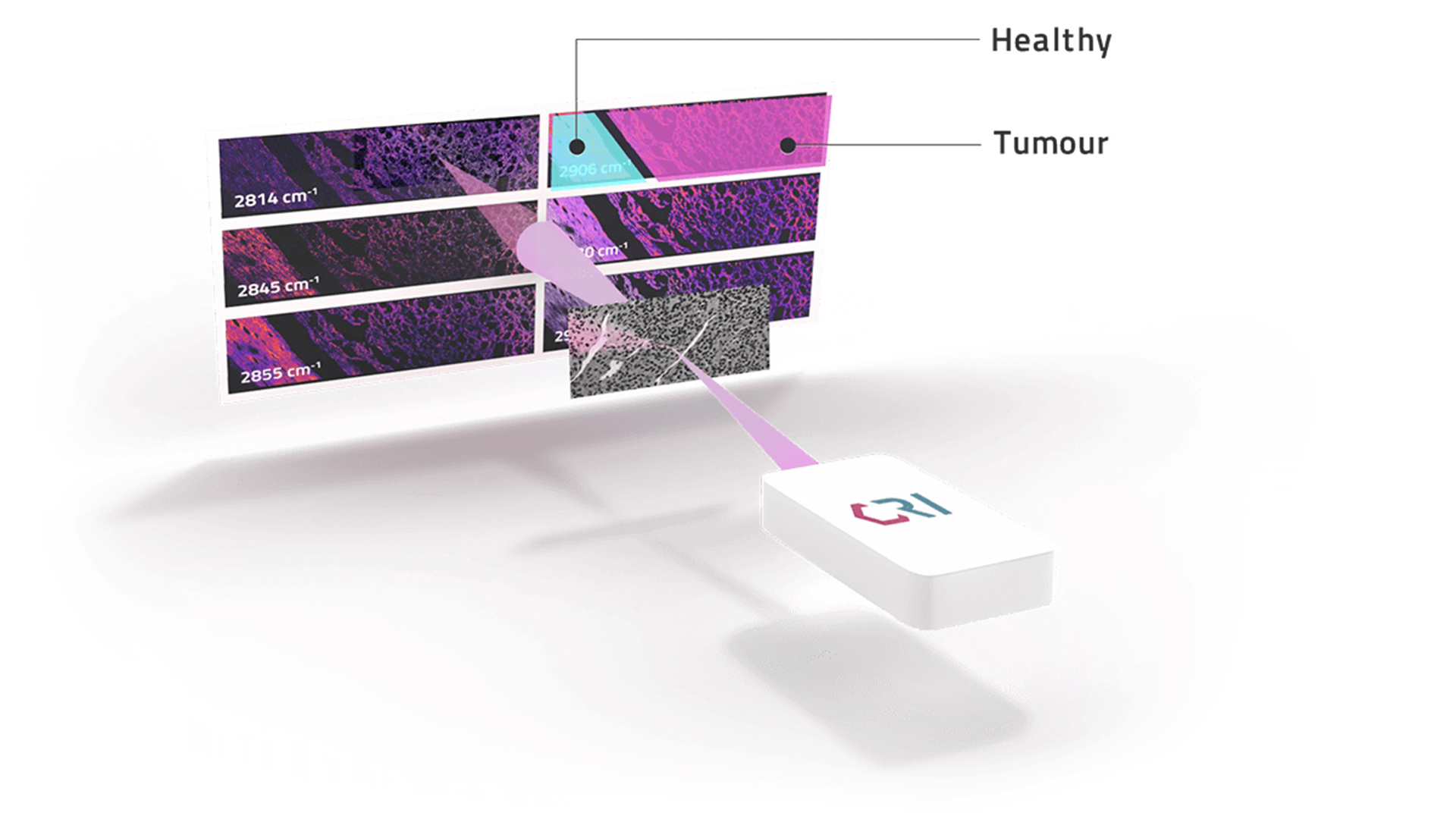
Graphene Flagship partner Cambridge Raman Imaging Limited (CRI) is leading the CHARM project with the aim of overcoming the limitations of the current methods used in cancer diagnosis. CRI is a spin-off of the Polytechnic University of Milan (Italy) and the University of Cambridge (UK).
Traditionally, cancer patients’ biopsies are stained with the hematoxylin and eosin (H&E) method: hematoxylin stains the nucleus while eosin stains the cytoplasm of the cells. This method allows the pathologist to visualise cellular structures, identify abnormal cell morphology, assess tissue architecture and detect any signs of cancerous growth. However, the method cannot measure molecular composition, is time consuming, and introduces variability between samples.
Using graphene-based biomedical technologies, the CHARM approach allows to save time and effort, as it can provide results in a matter of minutes. It eliminates the need for staining and enables the measurement of tissue molecular composition, which is crucial for determining tumour subtypes and grades. The technology takes advantage of coherent Raman scattering to measure the vibrational modes associated with chemical bonds present in the sample. AI-based methods elaborate the data to emulate the staining process and support pathologists in differentiating between normal and tumour tissues. Currently, the team is focusing on head and neck cancer as a representative case.
References
- Masvidal-Codina, Eduard, et al. “Characterization of optogenetically-induced cortical spreading depression in awake mice using graphene micro-transistor arrays.” Journal of Neural Engineering 18.5 (2021): 055002. https://iopscience.iop.org/article/10.1088/1741-2552/abecf3/meta
- Bonaccini Calia, Andrea, et al. “Full-bandwidth electrophysiology of seizures and epileptiform activity enabled by flexible graphene microtransistor depth neural probes.” Nature Nanotechnology 17.3 (2022): 301-309. https://www.nature.com/articles/s41565-021-01041-9
- Masvidal-Codina, Eduard, et al. “High-resolution mapping of infraslow cortical brain activity enabled by graphene microtransistors.” Nature Materials 18.3 (2019): 280-288. https://www.nature.com/articles/s41563-018-0249-4
- de la Cruz, Jose, et al. “Single and Multisite Graphene‐Based Electroretinography Recording Electrodes: A Benchmarking Study.” Advanced Materials Technologies 7.6 (2022): 2101181. https://onlinelibrary.wiley.com/doi/full/10.1002/admt.202101181
Please note, this article will also appear in the fifteenth edition of our quarterly publication.