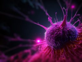
Chalmers University of Technology has unveiled a groundbreaking computer model capable of identifying signs of lymphatic cancer with an impressive accuracy rate of 90%.
In recent years, the field of medical image analysis has undergone a revolution with the integration of artificial intelligence (AI). This transformative technology has now reached a significant milestone in the realm of lymphoma detection.
The emergence of AI-driven methodologies for interpreting medical images holds promise across various medical domains.
These innovations not only alleviate the burden on radiologists by providing secondary opinions but also prioritise patient care by identifying cases requiring urgent attention.
Ida Häggström, Associate Professor at Chalmers’ Department of Electrical Engineering, explained: “An AI-based computer system for interpreting medical images also contributes to increased equality in healthcare by giving patients access to the same expertise and being able to have their images reviewed within a reasonable time, regardless of which hospital they are in.
“Since an AI system has access to much more information, it also makes it easier in rare diseases where radiologists rarely see images.”
Training the AI Model
The developed computer model, named Lars (Lymphoma Artificial Reader System), employs deep learning techniques to analyse positron emission tomography (PET) images.
The model’s training process involves supervised learning, where the computer evaluates images alongside their corresponding diagnoses to refine its predictive capabilities.
The AI model operates autonomously, discerning image patterns crucial for accurate predictions without pre-programmed instructions.
“I have used what is known as supervised training, where images are shown to the computer model, which then assesses whether the patient has lymphoma or not.
“The model also gets to see the true diagnosis, so if the assessment is wrong, the computer model is adjusted so that it gradually gets better and better at determining the diagnosis,” added Häggström.
A landmark study in lymphatic cancer detection
The recent study marks a significant stride in lymphoma diagnosis. Drawing from a vast dataset of over 17,000 images sourced from more than 5,000 lymphatic cancer patients, the team trained the computer model to identify visual cues indicative of lymph node cancer.
In their investigation, the researchers meticulously reviewed image archives spanning more than a decade. They meticulously compared the final diagnoses of patients with scans obtained from PET and computed tomography (CT) before and after treatment.
This comprehensive analysis formed the basis for training the AI computer model to identify signs indicative of lymphatic cancer in the images with 90% accuracy.
Challenges and future prospects
Despite achieving a commendable accuracy rate, Häggström acknowledges the need for extensive validation before integrating the model into clinical practice.
Challenges such as procuring ample image data and distinguishing cancerous features from treatment-induced changes underscore the complexity of AI-driven diagnostics.
“We have made the computer code available now so that other researchers can continue to work on the basis of our computer model, but the clinical tests that need to be done are extensive,” said Häggström.
The integration of AI in medical imaging heralds a new era in disease detection and diagnosis.
While significant strides have been made, ongoing validation and refinement are imperative to realise the full potential of AI in clinical settings.