Research led by a team of cancer researchers at the University of Cambridge, UK, suggests that a new advanced computing technique, which uses routine medical scans, can allow doctors to take more accurate tumour biopsies.
According to a paper published by the team in European Radiology, this is an important step towards precision tissue sampling for cancer patients to help select the best treatment. In the future, this technique could even replace clinical tumour biopsies with ‘virtual biopsies’, sparing patients invasive procedures.
Many cancer patients undergo several biopsies to confirm their diagnosis and plan their treatment. But because this is an invasive clinical procedure, there is an urgent need to reduce the number of biopsies taken and to make sure biopsies accurately sample the genetically-different cells in the tumour, particularly for ovarian cancer patients.
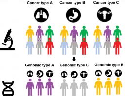
High grade serous ovarian (HGSO) cancer is referred to as a ‘silent killer’ because early symptoms can be difficult to identify. By the time the cancer is diagnosed, it is often at an advanced stage, and survival rates have not changed much over the last 20 years. HGSO tumours tend to have a high number of different types of cancer cells within the tumours, known as tumour heterogeneity, and patients with more tumour heterogeneity tend to have a poorer response to treatment.
Using high-powered computing methods to advise tumour biopsies
For the study, the patients first had a standard-of-care CT scan. The researchers then used a process called radiomics to identify and map distinct areas and features of the tumour using the data from the CT scan. The tumour map was then superimposed on the ultrasound image of the tumour and the combined image were used to guide the biopsy procedure.
By conducting targeted biopsies using this method, the research team reported that the diversity of cancer cells within the tumour was successfully captured.
Co-first author Dr Lucian Beer, from the Department of Radiology and CRUK Cambridge Centre Ovarian Cancer Programme, said of the results: “Our study is a step forward to non-invasively unravel tumour heterogeneity by using standard-of-care CT-based radiomic tumour habitats for ultrasound-guided targeted biopsies.”